| OBSERVATION
The retina is thinnest at the fovea centralis, the center of the macula. The reitna is normal transparent, and some of the incident light is reflected at the vitroretinal interface. The resulting sheen is especially noticable when young heavily pigmented patients are examined with an indirect ophthalmoscope. Absence of this foveal reflection may indicate disease, but the reflection may be absent in blond or elderly patients even though the retina is normal.
ANATOMY The fovea centralis, which lies about 3.5mm lateral to the optic disc, is specialised for fine visual discrimination. In the fovea, the receptors are all cones; the outer nuclear layer is thinned; the other parenchymal layers are displaced centrifugally; and the internal limiting membrane is thin. Throughtout most of the retina, the axons of the receptor cells pass directly to the inner side of the outer plexiform layer, where they connect with dendrites of horizontal and bipolar cells, which extend outward from the inner nuclear layer. In the macula, however, the receptor cell axons ollow an oblique course and are called the Henle fibre layer. The normally empty extracellular space of the retina is potentially greatest at the macula, and diseases that lead to accumulation of extracellular material cause considerable thickening in this area.
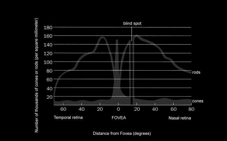
SCIENCE The cones are used for detailed vision and color perception. They predominate at the macula, the center of visual attention; and they alone are present at the fovea, the site of optimal visual acuity. The rods, which predominate elsewhere, function best in reduced illumination. The principle roles of the extracellular retina are night vision and visual orientation. |